
Pathology Case of the Month - Raccoon
Case History: An adult female 3.39 kg Common Raccoon was found dead at a private residence in Wisconsin, USA. The raccoon was collected for evaluation following observations by the landowner of 2 dead and 3 sick raccoons over the previous week. Symptoms observed in sick animals included walking slowly, back legs giving out, and lethargy. Affected animals were approachable and/or unresponsive.
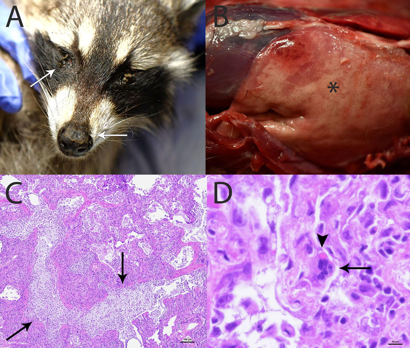
Gross Findings: On external examination, there is green-gray mucoid discharge around both eyes and crusting on the surface of the nose. (Fig 1A). On internal examination, there is adequate subcutaneous, visceral, and epicardial fat. The lungs fail to collapse on opening of the thoracic cavity. Rib impressions are present on the surface of the right lung lobes, which are deep red. The left cranial lung lobe is diffusely tan and consolidated (Fig 1B).
Histopathological Findings: There were minimal lesions in the brain, with rare, mild lymphocytic perivascular cuffing. Marked lymphoid depletion was seen in the spleen. In the lung, there was severe suppurative and histiocytic bronchointerstitial pneumonia (Fig 1C). Multinucleated syncytial cells were seen in many airways. Brightly eosinophilic, intranuclear and intracytoplasmic, variably sized viral inclusions were numerous throughout the section, within macrophages, syncytial cells, and epithelial cells. (1D).
Morphologic Diagnosis: Acute to subacute, severe, multifocal to coalescing suppurative and histiocytic bronchointerstitial pneumonia with intralesional viral inclusions.
Disease: Canine Distemper
Etiology: Canine Distemper Virus (CDV). CDV is a member of the genus Morbillivirus in the family Paramyxoviridae.
Distribution: Worldwide
Seasonality: Any time of year, but in raccoons in North America, there is increased prevalence during mating season in the spring, when there is high intraspecific contact and an increase in susceptible juveniles.
Host range: Well-known historically as a cause of often fatal disease in domestic dogs, CDV has since been found to infect all families of terrestrial carnivores: Canidae, Felidae, Hyaenidae, Mustelidae, Procyonidae, Ursidae, and Viverridae. Disease develops most commonly in young or immunocompromised individuals.
Transmission: CDV is highly contagious. It is shed in secretions and excretions (urine, feces, ocular and nasal discharges) of infected animals, with transmission primarily by aerosolization.
Clinical signs: Acute systemic illness is typically characterized by multiple clinical signs, including mucopurulent oculonasal discharge, cough, depression, anorexia, vomiting, and diarrhea. In animals that survive the initial systemic disease, neurologic signs may develop, with seizures, ataxia, paresis, and myoclonus being the most common. Neurologic signs may be accompanied by digital hyperkeratosis (“hard pads”).
Pathology: Pneumonia and catarrhal enteritis are hallmarks of acute systemic infection, along with lymphoid depletion. Pneumonia is diffuse and interstitial; secondary bacterial infections are common. Syncytial cells are often present. CDV inclusions are found in epithelial cells, especially in the lungs, bile ducts, and urinary bladder, as well as in neurons, glia, and leukocytes. Inclusions are eosinophilic, 1 to 5 µm in diameter, and can be intracytoplasmic or intranuclear. Neonatal animals may develop primary fatal CNS demyelination; older animals with neurologic involvement develop encephalomyelitis, characterized by widespread perivascular lymphoplasmacytic infiltrates, multifocal neuronal degeneration, as well as areas of demyelination.
Diagnosis: A presumptive diagnosis can be made on a live animal based on clinical signs, however a definitive diagnosis requires histopathologic and/or laboratory testing. Wild animals with neurologic signs must be considered rabies suspects until proven otherwise and should be euthanized and screened for rabies. A definitive diagnosis of CDV in a dead animal should be made based upon characteristic gross and histopathologic findings plus a positive PCR test or immunohistochemistry.
Public health concerns: There are no known reports of CDV infecting humans. However, canine distemper has been reported in non-human primates [Rhesus Monkey (Macaca mulatta) and Cynomolgus Macaques (Macaca fascicularis)] with high mortality rates. Infections in these primates have raised concerns that CDV could have the potential to become a human pathogen in the future.
Wildlife population impacts: Given this virus’s wide range of hosts and ease of transmission, local decreases in some species may be caused when an outbreak occurs. Populations of endangered species, such as the Black Footed Ferret (Mustela nigripes), may be devastated by this disease.
Management: Vaccination is very effective in preventing disease. Sick and dead animals should be removed from the population when an outbreak occurs. Routine disinfection procedures are usually effective in destroying CDV in kennels or rehab facilities.
References:
- Deem SL, Spelman LH, Yates RA, Montali RJ. Canine distemper in terrestrial carnivores: a review. J Zoo Wildl Med. 2000 31:441-51. https://doi.org/10.1638/1042-7260(2000)031[0441:cditca]2.0.co;2
- Giacinti JA, Pearl DL, Ojkic D, Bondo K, Jardine CM. 2023. Canine distemper virus ecology: Insights from a longitudinal serologic study in wild raccoons (Procyon lotor). J Wildl Dis. 159:407-419. https://doi.org/10.7589/JWD-D-22-00052
- Loots AK, Mitchell E, Dalton DL, Kotzé A, Venter EH. 2017. Advances in canine distemper virus pathogenesis research: a wildlife perspective. J Gen Virol. 98:311-321. https://doi.org/10.1099/jgv.0.000666
- Nemeth MN, Yabsley MJ. 2021. SCWDS Field Manual of Wildlife Diseases in the Southeastern United States, 4th Edition. Southeastern Cooperative Wildlife Disease Study, Athens.
- Roelofs D, Schmitz KS, van Amerongen G, Rijsbergen LC, Laksono BM, Comvalius AD, Nambulli S, Rennick LJ, van Run P, Duprex WP, van den Brand JMA, de Swart RL, de Vries RD. 2023. Inoculation of raccoons with a wild-type-based recombinant canine distemper virus results in viremia, lymphopenia, fever, and widespread histological lesions. mSphere 8:e00144-23. https://doi.org/10.1128/msphere.00144-23
Related Content

WHISPers

Pathology Case of the Month

Diagnostic Services
Related Content
WHISPers
Pathology Case of the Month
Diagnostic Services
Get Our News
These items are in the RSS feed format (Really Simple Syndication) based on categories such as topics, locations, and more. You can install and RSS reader browser extension, software, or use a third-party service to receive immediate news updates depending on the feed that you have added. If you click the feed links below, they may look strange because they are simply XML code. An RSS reader can easily read this code and push out a notification to you when something new is posted to our site.