
Pathology Case of the Month - Canada Geese
Case History: In October of 2022, a morbidity and mortality event in Canada Geese (Branta canadensis) was reported at a retention pond in Wisconsin, USA. The estimated number of affected geese at the time of submission was 21 dead and 12 sick. Two geese were found dead and submitted for diagnostic necropsy.
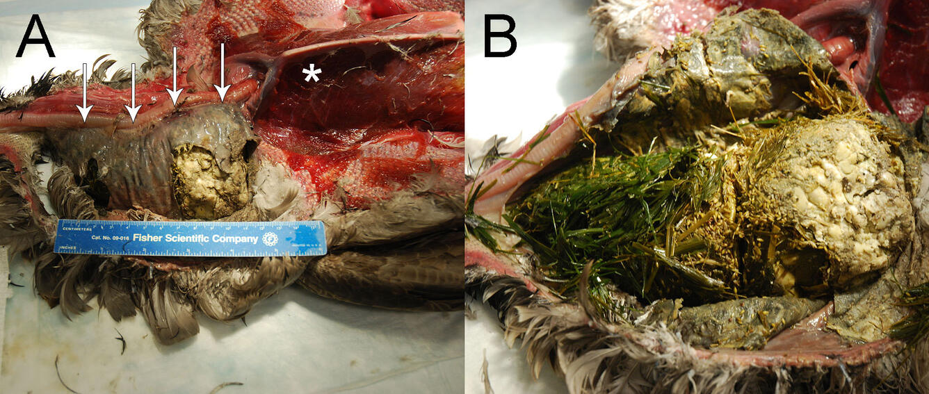
Gross Findings: Two juvenile Canada Geese (one male, one female) in emaciated body condition and fair postmortem condition were examined at necropsy and had similar findings. The pectoral muscles were severely thin (Fig. 1A), and there were no subcutaneous, visceral, or epicardial fat stores. In the ventral neck region just in front of the thoracic inlet there was a doughy mass ranging from ~12 x 4 x 4 cm to ~18 x 8 x 8.5 cm. The esophagus in this region was markedly distended by impacted ingesta (Fig. 1A) composed of (from proximal to distal) abundant grass, abundant small (2 x 1 mm) pale yellow seeds, and abundant halved, white, kidney bean-shaped legumes (soybeans) (Fig. 1B). One bird had fewer soybeans but had a moderate number of kernels of corn. The underlying esophageal mucosa was mildly thickened, roughened, and green grey with areas of yellow. The liver was very small, only extending ~1 cm over the ventriculus. The kidneys were also subjectively small. All other tissues were within normal limits.
Histopathological Findings: In one bird examined microscopically, the esophagus proximal to the impaction site is unremarkable, with normal wall layering and the presence of submucosal glands (Fig. 2A). The esophagus from the impacted area has changes consistent with pressure necrosis (Fig. 2B). The esophageal mucosa is multifocally ulcerated, with loss of epithelial cells and replacement by fibrin or rare clusters of degenerate inflammatory cells (presumed heterophils). The submucosa and serosa are markedly thickened by streams of fibroblasts and small amounts of collagen (early fibrosis) with small numbers of heterophils superficially beneath the areas of ulceration. The submucosal glands are absent. The tunica muscularis is thin and partially replaced by similar fibrous tissue, and in one region, there are increased amounts of collagen and perpendicularly arranged blood vessels that completely replace the tunica muscularis. Artifactually separated from the mucosa are serocellular crusts composed of abundant eosinophilic cellular and karyorrhectic nuclear debris admixed with many degenerate inflammatory cells (presumed heterophils) and various amounts of plant material, multifocal mixed colonies of gram-positive and gram-negative bacteria, and a small number of fungal hyphae. None of these fungal and bacterial organisms are apparent in the parenchyma of the esophagus. Other microscopic findings include severe atrophy of fat stores and moderate to severe atrophy of skeletal muscles (consistent with emaciation), moderate numbers of eosinophilic spherules in the lumina of the ureters (suspect secondary to dehydration), and moderate hemosiderosis in the liver and spleen (incidental findings).

Diagnostic Test Results: Both geese had similar test results. Liver lead screening was in the low-level background range (<3.0 ppm dry weight). Brain cholinesterase activity was not inhibited, indicating that exposure to organophosphate or carbamate pesticides was unlikely. Routine and fungal culture of the esophagus and esophageal content had heavy growth of mixed gram-positive and gram-negative bacteria, yeast, and fungal species interpreted as secondary infections due to the impaction. Tracheal and cloacal swabs were negative for avian influenza by matrix RT-PCR.
Morphologic Diagnoses:
- Esophageal impaction by soybeans
- Emaciation
Condition: Esophageal impaction
Etiology: In this case, esophageal impaction was due to ingestion of soybeans. Other causes of esophageal impaction include ingestion of other dry plant materials (i.e., cowpeas, green plants), excess feed intake, heavy infection with ventricular nematodes (Amidostomum sp.), and ingestion of fishing sinkers or lead shotgun pellets causing a mechanical obstruction or leading to lead poisoning. Occasionally, upper gastrointestinal impaction is observed with aquatic bird bornavirus 1 infection.
Distribution: Worldwide, wherever there is exposure to soybeans.
Seasonality: Early fall to winter, centering around soybean harvest time.
Host range: Esophageal impaction by soybeans has been frequently reported in free-ranging Canada Geese in North America. Other Anatidae (ducks, geese, and swans) species that also feed on soybeans may be affected.
Pathogenesis: Geese feed on soybeans that have been shattered during the harvesting process. The soybeans must be dry to cause an impaction. After dry soybeans are ingested and the bird drinks water, the soybean swell, leading to obstruction and pressure necrosis of the esophagus. In one report, soybeans were found to expand two and one-half times their previous volume after exposure to water.
Other factors: Precipitation, harvesting schedules, and migration patterns affect the extent of mortality from soybean impaction. When precipitation preceding soybean harvesting is lower than expected, mortalities are increased as shattered soybeans are more likely to be dry. Geese will preferentially feed on corn, so the harvest schedule of corn also plays a part, with early and rapid corn harvesting decreasing mortality caused by soybean impaction. Early migration of birds in the fall will increase the length of time that geese can feed on soybeans, increasing the number of birds that can become impacted.
Clinical signs: Soybean impaction occurs in two forms. In the acute form, geese die within 24 hours of soybean ingestion with little to no clinical signs of impaction. In the chronic form, geese are unable to extend their necks and will hold their heads towards the body, forming a curve in the neck. Birds will isolate themselves and often move very little unless disturbed. Geese may continue to try to drink and eat for a time (leading to the presence of other food elements in the impacted material), but eventually the inability to take in sustenance leads to emaciation and death due to starvation.
Pathology: On gross examination, geese with the acute form have normal body weight and fat stores. The mid-cervical to thoracic esophagus is distended with fresh, swollen soybeans. Occasionally the esophagus may be ruptured. In the chronic form, usually only the cervical portion of the esophagus is involved, and there is severe pressure necrosis of the esophagus, often with blackening and lichenification of the wall. Affected birds are emaciated, with loss of muscle mass and fat stores. Microscopically, there can be changes in the affected esophagus consistent with pressure necrosis, including ulceration and varying amounts of fibrosis expanding and effacing the wall.
Diagnosis: A diagnosis can be made based on clinical signs and gross findings of an esophagus impacted with soybeans with (acute form) or without (chronic form) pressure necrosis. Additional diagnostic testing should be performed to rule out lead toxicosis or bornavirus 1 infection as a cause of esophageal impaction, or other acute poisoning agent that may cause sudden death.
Public health concerns: None
Wildlife population impacts: The number of affected geese can vary considerably between years, depending on the amount of rainfall and harvest times of soybeans and corn. In one report from a wildlife refuge in Illinois, mortalities from soybean impaction ranged from less than 1% to almost 20% of the goose population.
Management: Mitigation attempts can be used to reduce the number of mortalities from soybean impaction and include reducing the availability of dry soybeans by disking or plowing under the shattered soybeans immediately after they are harvested, hazing the birds from the fields, or artificially supplying an alternate food source such as corn (if the corn harvest is delayed or slowed). Non-surgical and surgical treatments in individual geese have been reported, including administration of subcutaneous fluid, external palpation to manipulate the esophageal contents up to the mouth, esophageal gavage, and esophagotomy.
References:
- Durant AJ. 1956. Impaction and pressure necrosis in Canada geese due to eating dry hulled soybeans. J Wildl Manag 20(4):399-404. https://www.jstor.org/stable/3797151
- Fenton H, McManamon R, Howerth EW. Anserioformes, Ciconiiformes, Charadriiformes, and Gruiformes. In: Pathology of Wildlife and Zoo Animals. Terio KA, McAloose D, St. Leger J, editors. Elsevier, San Diego, CA, pp. 697-721. http://dx.doi.org/10.1016/B978-0-12-805306-5.00029-8
- Hansen HA, McNeil CW, Priebe MD. 1957. Mortality of Canada geese with impacted gullets in Eastern Washington, 1949-1954. J Wildl Manag 21(1):96-98. https://www.jstor.org/stable/3797691
- Jarvis RL. 1976. Soybean impaction in Canada geese. Wildl Soc Bull 4(4):175-179. https://www.jstor.org/stable/3781925
- MacNeill AC and Barnard T. 1978. Necropsy results in free-flying and captive Anatidae in British Columbia. Can Vet J 19:17021. https://www.ncbi.nlm.nih.gov/pmc/articles/PMC1789336/
- Muscatello G. 1998. Oesphageal impaction in a Canada goose (Branta canadensis). Aust Vet J 76(8):537-540. https://doi.org/10.1111/j.1751-0813.1998.tb10209.x
Related Content

WHISPers

Pathology Case of the Month

Diagnostic Services
Related Content
WHISPers
Pathology Case of the Month
Diagnostic Services
Get Our News
These items are in the RSS feed format (Really Simple Syndication) based on categories such as topics, locations, and more. You can install and RSS reader browser extension, software, or use a third-party service to receive immediate news updates depending on the feed that you have added. If you click the feed links below, they may look strange because they are simply XML code. An RSS reader can easily read this code and push out a notification to you when something new is posted to our site.