
Histology panel from eastern gray squirrel

Detailed Description
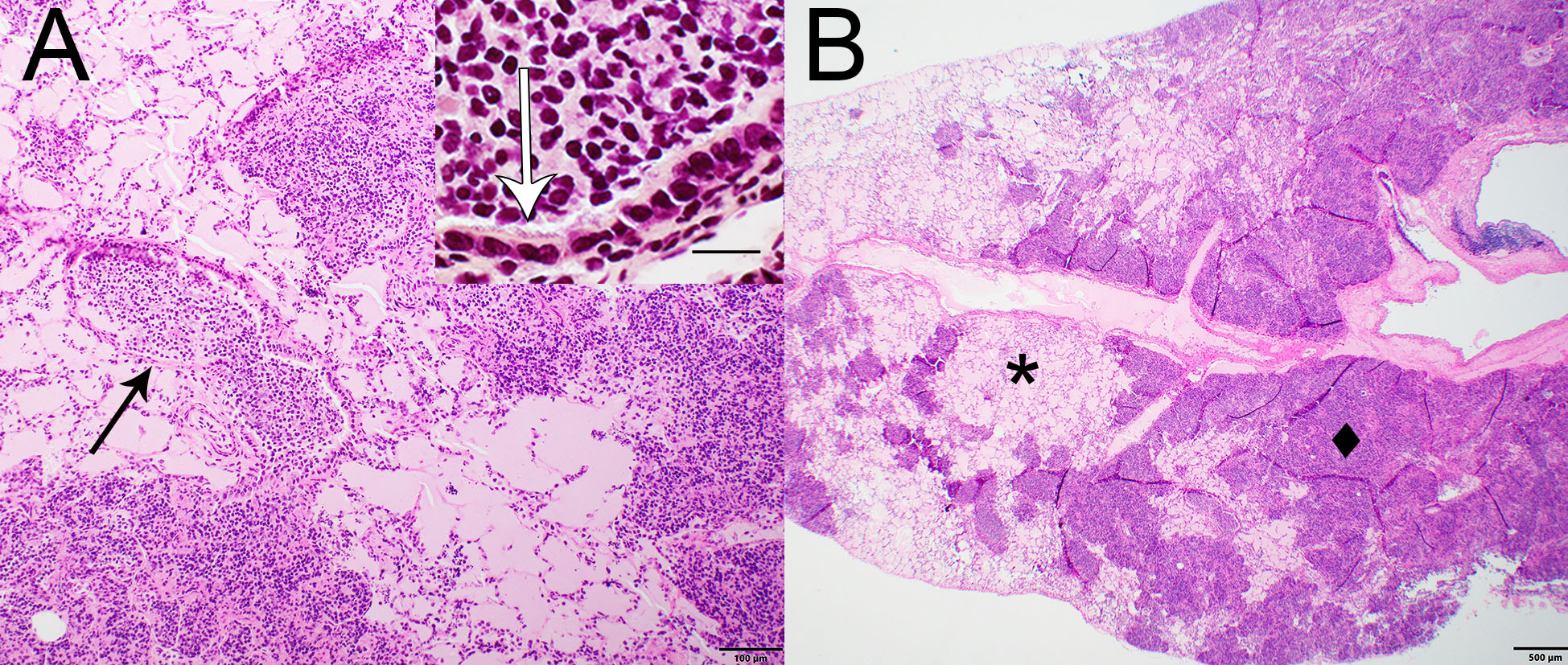
Photomicrographs from the lung of an eastern gray squirrel (Sciurus carolinensis) found dead in Wisconsin, U.S.A. (A) A bronchiole (arrow) contains numerous neutrophils. H&E stain. Inset: Bronchiolar epithelium is overlain by Gram-negative bacteria. Brown and Hopps stain. Bar = 20 µm. (B) Approximately 30% of the pulmonary parenchyma is replaced by dense basophilic aggregates of inflammatory cells (diamond); the hilar region is most affected. Remaining alveolar spaces contain pale eosinophilic fluid (edema; asterisk). H&E stain.
Sources/Usage
Public Domain.