
Photomicrograph from a raccoon found sick and euthanized in Texas

Detailed Description
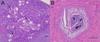
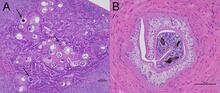
Photomicrograph from a raccoon (Procyon lotor) found sick and euthanized in Texas, US. (A) A cluster of schistosome ova are present in the pancreas. Ova are thin-shelled and are not operculated. Each viable egg contains a miracidium (arrows). The ova are surrounded by infiltrates of eosinophils, macrophages, and lymphocytes. H&E stain. (B) Adult schistosomes in copula in a mesenteric blood vessel in a raccoon (Procyon lotor). Adults have no body cavity and are filled with parenchyma. Cecae containing dark brown pigmented material resulting from the breakdown of red blood cells can be seen. H&E stain.
Sources/Usage
Public Domain.