
Photomicrographs of lung from a mallard duck

Detailed Description
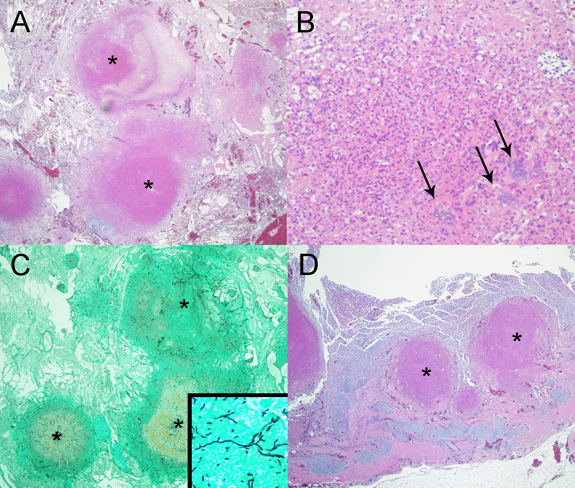
Photomicrographs from a mallard duck (Anas platyrhynchos) found dead in Idaho, USA. (A) Multiple granulomas (*) with eosinophilic necrotic centers efface the lung. H&E stain. (B) Granulomas contain many degenerate heterophils and a few multinucleated giant cells (arrows). H&E stain. (C) Granulomas (*) in the lung contain numerous fungal hyphae. Inset: Fungal hyphae have 3-8 µm-wide, thin parallel walls with regular septations and acute angle branching. GMS stain. (D) Multiple granulomas (*) similar to those in the lung efface the wall of the intestine. H&E stain.
Sources/Usage
Public Domain.