
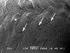
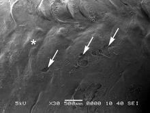
Figure 4. Scanning Electron Micrograph of Descaling Area

Detailed Description
Figure 4. Scanning electron micrograph of descaling area delimited by box in Figure 3 showing epidermal disruption (arrows), empty scale pockets and restoration of epidermal integrity (asterisk). An exposed scale with visible concentric ridges is visible at the lower center. Scale bar = 500 µm.
Sources/Usage
Public Domain.