
Images of heart, pancreas, lung, and brain tissue from gyrfalcon
Detailed Description
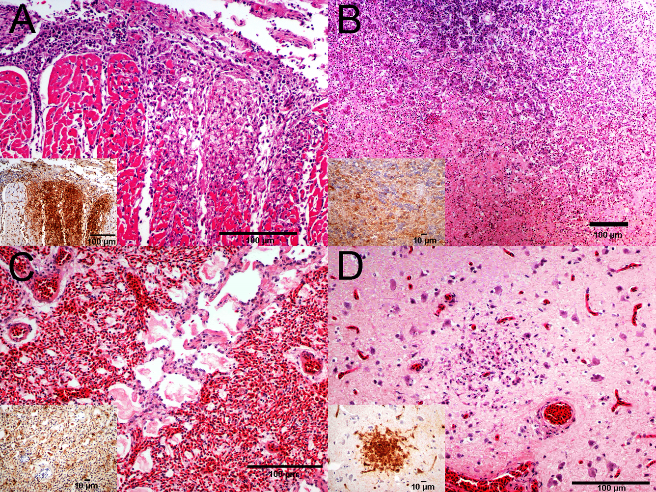
A. Heart, multifocal and focally extensive necrotizing myocarditis. H&E. Inset: Section of same heart tissue; immunohistochemical staining of influenza A virus antigen is strongly positive in areas of necrosis. B. Pancreas, multifocal and focally extensive necrosis of the acinar epithelium. H&E. Inset: Immunohistochemical staining of influenza A virus antigen is strongly positive in areas of necrosis. C. Lung, diffuse pulmonary congestion and edema. Hematoxylin and eosin (H&E) stain. Inset: Positive immunohistochemical staining of influenza A virus antigen is present within pulmonary histiocytes and the capillary endothelium. D. Brain (cerebrum), focal necrotizing encephalitis. H&E. Inset: Areas of necrosis shows strong positive immunohistochemical staining of influenza A virus antigen.
Sources/Usage
Public Domain.