
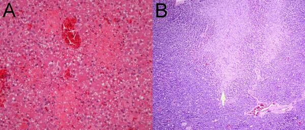
Photographs from an adult female mountain cottontail rabbit (Sylvilagus nuttallii) found dead in Montana, U.S.A. (A) Liver with random foci of necrosis (asterisk) characterized by accumulation of cellular detritus intermingled with fibrin and colonies of small coccoid bacteria (inset). H&E stain.
Images
Images from the National Wildlife Health Center.
Filter Total Items: 201

Photographs from organs of a mountain cottontail rabbit found dead
Photographs from an adult female mountain cottontail rabbit (Sylvilagus nuttallii) found dead in Montana, U.S.A. (A) Liver with random foci of necrosis (asterisk) characterized by accumulation of cellular detritus intermingled with fibrin and colonies of small coccoid bacteria (inset). H&E stain.

Photographs of intestine tissue from mallard ducks with trematodiasis
Photographs from two mallards (Anas platyrhynchos) found dead within a warm-water retention pond in Minnesota, US. (A) Caeca with mucosal roughening and caseous plaque formation. A larger trematode (arrow) is found within. (B) Dilated and thinned intestine containing watery intestinal content with pinpoint white material.
Photographs from two mallards (Anas platyrhynchos) found dead within a warm-water retention pond in Minnesota, US. (A) Caeca with mucosal roughening and caseous plaque formation. A larger trematode (arrow) is found within. (B) Dilated and thinned intestine containing watery intestinal content with pinpoint white material.

Photographs from two mallards (Anas platyrhynchos) with trematodiasis
Photographs from two mallards (Anas platyrhynchos) found dead within a warm-water retention pond in Minnesota, US. (A) Jejunum with superficial pseudomembranes and deep mucosal granulomatous inflammation (clear arrow). Small degenerated metazoan parasites are present rarely within the inflamed areas (black arrow). H&E stain. (B).
Photographs from two mallards (Anas platyrhynchos) found dead within a warm-water retention pond in Minnesota, US. (A) Jejunum with superficial pseudomembranes and deep mucosal granulomatous inflammation (clear arrow). Small degenerated metazoan parasites are present rarely within the inflamed areas (black arrow). H&E stain. (B).

Photographs from a Common Eider (Somateria mollissima)
Photographs from a Common Eider (Somateria mollissima) from Massachusetts, USA. (A) There are scattered, small, pale foci in the liver. (B) Petechial and ecchymotic hemorrhages are present in the pancreas.
Photographs from a Common Eider (Somateria mollissima) from Massachusetts, USA. (A) There are scattered, small, pale foci in the liver. (B) Petechial and ecchymotic hemorrhages are present in the pancreas.
Photomicrographs from a Common Eider (Somateria mollissima)
Photomicrographs from a Common Eider (Somateria mollissima) from Massachusetts, USA. (A) Multiple pale foci of acute hepatic necrosis. (B) Multiple foci of acute pancreatic necrosis (arrow).
Photomicrographs from a Common Eider (Somateria mollissima) from Massachusetts, USA. (A) Multiple pale foci of acute hepatic necrosis. (B) Multiple foci of acute pancreatic necrosis (arrow).

Pathologist examines a cottontail for cause-of-death determination
USGS National Wildlife Health Center pathologist examines a cottontail for cause-of-death determination.
USGS National Wildlife Health Center pathologist examines a cottontail for cause-of-death determination.

Eastern newt (Notophthalmus viridescens) in Wisconsin
Eastern newt (Notophthalmus viridescens) in Wisconsin.
Eastern newt (Notophthalmus viridescens) in Wisconsin.

American toad tucking into the leaf litter
American toad tucking into the leaf litter seen during fieldwork in Wisconsin.
American toad tucking into the leaf litter seen during fieldwork in Wisconsin.

Green frog in wetland vegetation
A green frog camouflaged in wetland vegetation that was seen during fieldwork in Wisconsin.
A green frog camouflaged in wetland vegetation that was seen during fieldwork in Wisconsin.

Cope’s grey treefrog
Cope’s grey treefrog on a tree trunk. Seen during fieldwork in Wisconsin.
Cope’s grey treefrog on a tree trunk. Seen during fieldwork in Wisconsin.

Cope’s grey treefrog, green morph
Cope’s grey treefrog, green morph, on a tree trunk. Seen during fieldwork in Wisconsin.
Cope’s grey treefrog, green morph, on a tree trunk. Seen during fieldwork in Wisconsin.

Images from necropsy of two foothill yellow-legged frogs (Rana boylii)
Two foothill yellow-legged frogs (Rana boylii) found dead in Santa Clara, California, USA. (A) One animal had pinpoint red foci on the ventral abdomen. (B) Another animal had a diffusely reddened kidney (arrow).
Two foothill yellow-legged frogs (Rana boylii) found dead in Santa Clara, California, USA. (A) One animal had pinpoint red foci on the ventral abdomen. (B) Another animal had a diffusely reddened kidney (arrow).

Photomicrographs from a foothill yellow-legged frog (Rana boylii)
Photomicrographs from a foothill yellow-legged frog (Rana boylii) found dead in Santa Clara, California, USA. (A) Small areas of epidermal necrosis with apoptotic keratinocytes and nuclear debris are multifocally present (arrow). (B) The liver shows randomly distributed, variably sized areas of coagulative necrosis (*).
Photomicrographs from a foothill yellow-legged frog (Rana boylii) found dead in Santa Clara, California, USA. (A) Small areas of epidermal necrosis with apoptotic keratinocytes and nuclear debris are multifocally present (arrow). (B) The liver shows randomly distributed, variably sized areas of coagulative necrosis (*).

Photograph from a muskrat (Ondatra zibethicus) found dead in Ohio, USA
Photograph from a muskrat (Ondatra zibethicus) found dead in Ohio, USA. The liver contained disseminated pinpoint to 1-mm diameter white foci (arrows). Tyzzer's disease.
Photograph from a muskrat (Ondatra zibethicus) found dead in Ohio, USA. The liver contained disseminated pinpoint to 1-mm diameter white foci (arrows). Tyzzer's disease.

Photographs from a muskrat (Ondatra zibethicus) liver
Photographs from a muskrat (Ondatra zibethicus) found dead in Ohio, USA. (A) Throughout the liver are random areas of hepatocellular necrosis (star). H&E stain. (B) Hepatocytes at the periphery of necrotic areas multifocally contain packets of argyrophilic bacterial rods (arrows). Modified Steiner’s stain.
Photographs from a muskrat (Ondatra zibethicus) found dead in Ohio, USA. (A) Throughout the liver are random areas of hepatocellular necrosis (star). H&E stain. (B) Hepatocytes at the periphery of necrotic areas multifocally contain packets of argyrophilic bacterial rods (arrows). Modified Steiner’s stain.

White-tailed deer in snow
A white-tailed deer (Odocoileus virginianus) in a snowy field in front of forest in Wisconsin.
A white-tailed deer (Odocoileus virginianus) in a snowy field in front of forest in Wisconsin.

A sea otter mom nursing her pup
A sea otter mom nursing her pup. Photo taken in Prince William Sound, Alaska. A new born sea otter needs to stay with its mother for six months to learn how to survive on its own.
A sea otter mom nursing her pup. Photo taken in Prince William Sound, Alaska. A new born sea otter needs to stay with its mother for six months to learn how to survive on its own.

Lung from a captive, adult male, 930-g Gyrfalcon, found dead in WA
Lung from a captive, adult male, 930-g Gyrfalcon, found dead in Washington, USA. Lung shows diffuse severe congestion and edema.
Lung from a captive, adult male, 930-g Gyrfalcon, found dead in Washington, USA. Lung shows diffuse severe congestion and edema.

Great-Horned Owl and Eurasian Collared-Dove found dead in Utah
Photographs from a Great-Horned Owl and Eurasian Collared-Dove found dead in a residential yard in Utah, U.S. (A) The owl had singeing of the rictal bristles on the right side of the face (inset), swollen right eyelids, and a cloudy and thickened right cornea.
Photographs from a Great-Horned Owl and Eurasian Collared-Dove found dead in a residential yard in Utah, U.S. (A) The owl had singeing of the rictal bristles on the right side of the face (inset), swollen right eyelids, and a cloudy and thickened right cornea.

Photomicrographs from a Great-Horned Owl
Photomicrographs from a Great-Horned Owl showing (A) moderate acute hemorrhage in the right atrial epicardium extending into the myocardium and (B) a focally extensive area of coagulative necrosis of the epidermis and dermis (arrow) consistent with an electrical burn.
Photomicrographs from a Great-Horned Owl showing (A) moderate acute hemorrhage in the right atrial epicardium extending into the myocardium and (B) a focally extensive area of coagulative necrosis of the epidermis and dermis (arrow) consistent with an electrical burn.

Histology of snake fungal disease
(A) Underneath the β-layer of the epidermis is an accumulation of hypereosinophilic necrotic debris. The remaining epidermis is extensively ulcerated.
(A) Underneath the β-layer of the epidermis is an accumulation of hypereosinophilic necrotic debris. The remaining epidermis is extensively ulcerated.