
Flushing stomach contents from a live resident Chinook salmon for an investigation of cannibalism and predation impacts.
Images
Search here for some of our available images.
Filter Total Items: 204

Flushing stomach contents from a live resident Chinook salmon
Flushing stomach contents from a live resident Chinook salmon for an investigation of cannibalism and predation impacts.
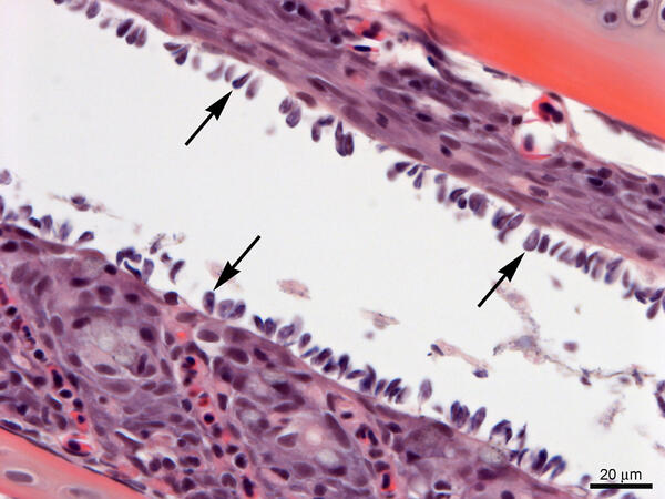
Gills from Lost River Suckers - heavy infestation of Ichthyobodo sp.
Gills from Lost River suckers with a heavy infestation of Ichthyobodo sp. (arrows). Slide is stained with hematoxylin and eosin.
Gills from Lost River suckers with a heavy infestation of Ichthyobodo sp. (arrows). Slide is stained with hematoxylin and eosin.

Herring (top) and juvenile Chinook salmon (bottom)
Herring (top) and juvenile Chinook salmon (bottom) flushed from the stomach of a resident Chinook salmon in Puget Sound.
Herring (top) and juvenile Chinook salmon (bottom) flushed from the stomach of a resident Chinook salmon in Puget Sound.

Nested polymerase chain reaction (nPCR) for the detection of Rs
Nested polymerase chain reaction (nPCR) for the detection of Renibacterium salmoninarum. Agarose gel electrophoresis is used for size separation and visualization of amplified DNA sequences.
Nested polymerase chain reaction (nPCR) for the detection of Renibacterium salmoninarum. Agarose gel electrophoresis is used for size separation and visualization of amplified DNA sequences.

Spawning Lost River suckers
Lost River suckers congregating to spawn on Sucker Springs in Upper Klamath Lake, Oregon.
Lost River suckers congregating to spawn on Sucker Springs in Upper Klamath Lake, Oregon.

WFRC nPCR test for detection of Renibacterium salmoninarum
Nested polymerase chain reaction (nPCR) for the detection of Renibacterium salmoninarum. Following two rounds of PCR amplification, samples are pipetted into an agarose gel for electrophoresis.
Nested polymerase chain reaction (nPCR) for the detection of Renibacterium salmoninarum. Following two rounds of PCR amplification, samples are pipetted into an agarose gel for electrophoresis.

WFRC nPCR test for detection of Renibacterium salmoninarum
Nested polymerase chain reaction (nPCR) for the detection of Renibacterium salmoninarum. Agarose gel electrophoresis is used for size separation and visualization of amplified DNA sequences.
Nested polymerase chain reaction (nPCR) for the detection of Renibacterium salmoninarum. Agarose gel electrophoresis is used for size separation and visualization of amplified DNA sequences.

Striped Bass diet sampling in the San Francisco Estuary
Sampling Striped Bass for diet items in the San Francisco Estuary.
Sampling Striped Bass for diet items in the San Francisco Estuary.

WFRC Quantitative Culture for Renibacterium salmoninarum Image 2
Spread plate procedure for quantitative culture Renibacterium salmoninarum from fish tissue or ovarian fluid.
Spread plate procedure for quantitative culture Renibacterium salmoninarum from fish tissue or ovarian fluid.

Willow Creek detection station PIT tag systems
Passive Integrated Transponder (PIT) tag detection station on Willow Creek, California. Systems like this one are used to detect movements of endangered Lost River and shortnose suckers in remote locations.
Passive Integrated Transponder (PIT) tag detection station on Willow Creek, California. Systems like this one are used to detect movements of endangered Lost River and shortnose suckers in remote locations.

Researcher works at microscope in Histology Lab
Tissue sections are mounted on glass slides, stained and examined with a microscope that magnifies cellular details up to 2,000 times with brightfield or fluorescence imaging. Microscopes are used in our research to understand the pathological changes caused by infectious agents such as bacteria, fungi, parasites and viruses.
Tissue sections are mounted on glass slides, stained and examined with a microscope that magnifies cellular details up to 2,000 times with brightfield or fluorescence imaging. Microscopes are used in our research to understand the pathological changes caused by infectious agents such as bacteria, fungi, parasites and viruses.

WFRC qPCR for detection of Renibacterium salmoninarum Image 1
Quantitative Polymerase Chain Reaction (qPCR) assay for the detection of Renibacterium salmoninarum DNA. A nucleic acid sequence detection system is shown.
Quantitative Polymerase Chain Reaction (qPCR) assay for the detection of Renibacterium salmoninarum DNA. A nucleic acid sequence detection system is shown.

WFRC qPCR for detection of Renibacterium salmoninarum Image 2
Quantitative polymerase chain reaction (qPCR) assay for the detection of Renibacterium salmoninarum DNA. Observation of qPCR results on the screen of a sequence detection system.
Quantitative polymerase chain reaction (qPCR) assay for the detection of Renibacterium salmoninarum DNA. Observation of qPCR results on the screen of a sequence detection system.

Hells Canyon on the Snake River
Hells Canyon on the Snake River.
Hells Canyon on the Snake River.

WFRC DFAT for detection of Renibacterium salmoninarum
Direct fluorescent antibody test (DFAT) for the detection of Renibacterium salmoninarum in tissues. Fluorescing R. salmoninarum cells are visible on a slide.
Direct fluorescent antibody test (DFAT) for the detection of Renibacterium salmoninarum in tissues. Fluorescing R. salmoninarum cells are visible on a slide.

Cle Elum River, a tributary of the Yakima River in Washington state
The Cle Elum River, a tributary of the Yakima River in Washington state. Sockeye salmon reintroduction efforts were initiated in the Cle Elum River by the Yakama Nation and Washington Department of Fish and Wildlife in 2009.
The Cle Elum River, a tributary of the Yakima River in Washington state. Sockeye salmon reintroduction efforts were initiated in the Cle Elum River by the Yakama Nation and Washington Department of Fish and Wildlife in 2009.

Sockeye salmon preparing to spawn
Sockeye salmon preparing to spawn upstream of Cle Elum Dam, Washington.
Sockeye salmon preparing to spawn upstream of Cle Elum Dam, Washington.

Howard Bay - blooms of cyanobacteria
As blooms of cyanobacteria die on the water surface of Upper Klamath Lake they turn blue-green.
As blooms of cyanobacteria die on the water surface of Upper Klamath Lake they turn blue-green.

In-lake mesocosms in Upper Klamath Lake, OR
USGS studies the behavior and health of juvenile endangered Lost River and shortnose suckers within in-lake mesocosms in Upper Klamath Lake, Oregon.
USGS studies the behavior and health of juvenile endangered Lost River and shortnose suckers within in-lake mesocosms in Upper Klamath Lake, Oregon.

Shoreline trap in Lookout Point Reservoir, OR
Shoreline traps in Lookout Point Reservoir, Oregon.
Shoreline traps in Lookout Point Reservoir, Oregon.

Juvenile Chinook salmon
Juvenile Chinook salmon on the Middle Fork Willamette River, Oregon.
Juvenile Chinook salmon on the Middle Fork Willamette River, Oregon.