
A giant garter snake (Thamnophis gigas) slithering through grass, photographed during field work in western Oregon.
Images
Images from the National Wildlife Health Center.
Filter Total Items: 200

Giant Garter Snake
A giant garter snake (Thamnophis gigas) slithering through grass, photographed during field work in western Oregon.

Chondroma mass on the hock of a sandhill crane
Photographs from a sandhill crane (Antigone canadensis) found dead in Indiana, U.S.A. (A) There is a firm mass on the right cranial hock with a roughened black and tan surface. (B) On cut section, the mass is gelatinous, mottled light pink to gray, and extends to the joint space (arrow).
Photographs from a sandhill crane (Antigone canadensis) found dead in Indiana, U.S.A. (A) There is a firm mass on the right cranial hock with a roughened black and tan surface. (B) On cut section, the mass is gelatinous, mottled light pink to gray, and extends to the joint space (arrow).

Photomicrographs from a sandhill crane found dead in Indiana U.S.A.
Photomicrographs from a sandhill crane (Antigone canadensis) found dead in Indiana, U.S.A. (A) The mass is composed of islands of well-differentiated chondrocytes separated by fibrovascular connective tissue. There is minimal cellular pleomorphism and no mitotic figures are seen (inset).
Photomicrographs from a sandhill crane (Antigone canadensis) found dead in Indiana, U.S.A. (A) The mass is composed of islands of well-differentiated chondrocytes separated by fibrovascular connective tissue. There is minimal cellular pleomorphism and no mitotic figures are seen (inset).
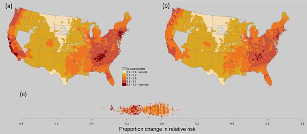
Maps of Bsal risk in the U.S. comparing pre- and post- action risk
Relative risk maps of combined Bsal risk, comparing (a) pre-action risk (2010–2015), to (b) risk after implementation of surveillance and of importation restrictions on over 200 salamander species. Relative risk scores were scaled to 2010–2015. (c) Change in relative risk score per county as proportion of pre-action risk.
Relative risk maps of combined Bsal risk, comparing (a) pre-action risk (2010–2015), to (b) risk after implementation of surveillance and of importation restrictions on over 200 salamander species. Relative risk scores were scaled to 2010–2015. (c) Change in relative risk score per county as proportion of pre-action risk.

Photomicrograph from a raccoon found sick and euthanized in Texas
Photomicrograph from a raccoon (Procyon lotor) found sick and euthanized in Texas, US. (A) A cluster of schistosome ova are present in the pancreas. Ova are thin-shelled and are not operculated. Each viable egg contains a miracidium (arrows). The ova are surrounded by infiltrates of eosinophils, macrophages, and lymphocytes. H&E stain.
Photomicrograph from a raccoon (Procyon lotor) found sick and euthanized in Texas, US. (A) A cluster of schistosome ova are present in the pancreas. Ova are thin-shelled and are not operculated. Each viable egg contains a miracidium (arrows). The ova are surrounded by infiltrates of eosinophils, macrophages, and lymphocytes. H&E stain.

Rat snake museum specimen
This rat snake is a preserved museum specimen with snake fungal disease that was collected in Tennessee in 1973. The photo was taken at the USGS National Wildlife Health Center in 2017 as part of a study.
This rat snake is a preserved museum specimen with snake fungal disease that was collected in Tennessee in 1973. The photo was taken at the USGS National Wildlife Health Center in 2017 as part of a study.

Nematodes in raccoon tissue
Photographs from a raccoon (Procyon lotor) from Arizona, US. (A) Large tan nematodes (arrowheads) are present within the subcutaneous tissue and muscle fascia in the distal right forelimb. (B) Closer view of the distal right forelimb showing the large tan nematodes (arrowheads).
Photographs from a raccoon (Procyon lotor) from Arizona, US. (A) Large tan nematodes (arrowheads) are present within the subcutaneous tissue and muscle fascia in the distal right forelimb. (B) Closer view of the distal right forelimb showing the large tan nematodes (arrowheads).

Photographs from a wild turkey found dead in Minnesota
Photographs from a wild turkey (Meleagris gallopavo) found dead in Minnesota, USA. (A) There are numerous multifocal to coalescing yellow proliferative lesions covering the eyelids, head, and neck with a region of ulceration and necrosis (*). (B) Yellow irregular plaques (arrow heads) multifocally cover the oral cavity.
Photographs from a wild turkey (Meleagris gallopavo) found dead in Minnesota, USA. (A) There are numerous multifocal to coalescing yellow proliferative lesions covering the eyelids, head, and neck with a region of ulceration and necrosis (*). (B) Yellow irregular plaques (arrow heads) multifocally cover the oral cavity.

Photomicrographs from a wild turkey found dead in Minnesota
Photomicrographs from a wild turkey (Meleagris gallopavo) found dead in Minnesota, USA. (A) The normal epithelium becomes markedly thickened due to hyperplasia of the stratum spinosum (left to right) with regions of ulceration and crusting (arrow). H&E stain.
Photomicrographs from a wild turkey (Meleagris gallopavo) found dead in Minnesota, USA. (A) The normal epithelium becomes markedly thickened due to hyperplasia of the stratum spinosum (left to right) with regions of ulceration and crusting (arrow). H&E stain.

Photograph & photomicrographs from exotic duck with avian tuberculosis
Photographs and photomicrographs from an exotic duck. (A) Grossly, the spleen is enlarged, firm, and yellow (arrow). Throughout the liver are multifocal to coalescing firm tan nodules (arrowheads). (B) The liver contains multifocal granulomas (asterisk). H&E stain.
Photographs and photomicrographs from an exotic duck. (A) Grossly, the spleen is enlarged, firm, and yellow (arrow). Throughout the liver are multifocal to coalescing firm tan nodules (arrowheads). (B) The liver contains multifocal granulomas (asterisk). H&E stain.

Photomicrographs from a big brown bat (Eptesicus fuscus) from Wisconsi
Photomicrographs from a big brown bat (Eptesicus fuscus) from Wisconsin, USA. H&E stain. (A) Low magnification of a cross section through the nares showing nasal cavity (*), oral mucosa (arrowhead), and haired skin (arrow). (B) Higher magnification showing a normal hair follicle (arrow) surrounded by sebaceous glands.
Photomicrographs from a big brown bat (Eptesicus fuscus) from Wisconsin, USA. H&E stain. (A) Low magnification of a cross section through the nares showing nasal cavity (*), oral mucosa (arrowhead), and haired skin (arrow). (B) Higher magnification showing a normal hair follicle (arrow) surrounded by sebaceous glands.

Photographs of lungs from a mallard duck found dead in Idaho
Photographs from a mallard duck (Anas platyrhynchos) found dead in Idaho, USA. (A) The lungs are dark red and have multifocal 1-3 mm diameter white to tan nodules disseminated throughout (arrows). (B) Cut section of the lung showing the nodules within the parenchyma (arrows).
Photographs from a mallard duck (Anas platyrhynchos) found dead in Idaho, USA. (A) The lungs are dark red and have multifocal 1-3 mm diameter white to tan nodules disseminated throughout (arrows). (B) Cut section of the lung showing the nodules within the parenchyma (arrows).

Photomicrographs of lung from a mallard duck
Photomicrographs from a mallard duck (Anas platyrhynchos) found dead in Idaho, USA. (A) Multiple granulomas (*) with eosinophilic necrotic centers efface the lung. H&E stain. (B) Granulomas contain many degenerate heterophils and a few multinucleated giant cells (arrows). H&E stain.
Photomicrographs from a mallard duck (Anas platyrhynchos) found dead in Idaho, USA. (A) Multiple granulomas (*) with eosinophilic necrotic centers efface the lung. H&E stain. (B) Granulomas contain many degenerate heterophils and a few multinucleated giant cells (arrows). H&E stain.

Administering the white-nose syndrome vaccine to a bat
Administering the white-nose syndrome vaccine to a bat during a field trial.
Administering the white-nose syndrome vaccine to a bat during a field trial.

Bat receiving white-nose syndrome vaccine
A bat receiving the white-nose syndrome vaccine during a field trial to study vaccine efficacy.
A bat receiving the white-nose syndrome vaccine during a field trial to study vaccine efficacy.

Cluster of cave myotis bats (Myotis velifer) on cave wall in Texas
Cluster of cave myotis bats (Myotis velifer) on cave wall in Texas.
Cluster of cave myotis bats (Myotis velifer) on cave wall in Texas.

Photographs from a red wolf (Canis rufus) found dead in North Carolina
Photographs from a red wolf (Canis rufus) found dead in North Carolina, USA. (A) Adult heartworms (Dirolfilaria immitis) in the right ventricle and atria of the heart and extending into the pulmonary artery. (B) Hard, haired nodule on the medial surface of the distal radius of the right leg.
Photographs from a red wolf (Canis rufus) found dead in North Carolina, USA. (A) Adult heartworms (Dirolfilaria immitis) in the right ventricle and atria of the heart and extending into the pulmonary artery. (B) Hard, haired nodule on the medial surface of the distal radius of the right leg.

Histology panel from eastern gray squirrel
Photomicrographs from the lung of an eastern gray squirrel (Sciurus carolinensis) found dead in Wisconsin, U.S.A. (A) A bronchiole (arrow) contains numerous neutrophils. H&E stain. Inset: Bronchiolar epithelium is overlain by Gram-negative bacteria. Brown and Hopps stain. Bar = 20 µm.
Photomicrographs from the lung of an eastern gray squirrel (Sciurus carolinensis) found dead in Wisconsin, U.S.A. (A) A bronchiole (arrow) contains numerous neutrophils. H&E stain. Inset: Bronchiolar epithelium is overlain by Gram-negative bacteria. Brown and Hopps stain. Bar = 20 µm.

Necropsy photos from eastern gray squirrel
Photographs from an eastern gray squirrel (Sciurus carolinensis) found dead in Wisconsin, U.S.A. (A) The lung contained multifocal firm red areas (arrows) and multifocal areas of hemorrhage (arrowhead). (B) Firm areas are red to tan on cut section and airways contain mucoid red to tan fluid.
Photographs from an eastern gray squirrel (Sciurus carolinensis) found dead in Wisconsin, U.S.A. (A) The lung contained multifocal firm red areas (arrows) and multifocal areas of hemorrhage (arrowhead). (B) Firm areas are red to tan on cut section and airways contain mucoid red to tan fluid.

Photomicrographs from a desert cottontail found dead in Texas
Photomicrographs from a desert cottontail found dead in Texas, U.S.A. showing (A) panlobular hepatocellular dissociation and necrosis and (B) intra-alveolar edema fluid (asterisk) and foamy pulmonary macrophages (arrows) in the lung.
Photomicrographs from a desert cottontail found dead in Texas, U.S.A. showing (A) panlobular hepatocellular dissociation and necrosis and (B) intra-alveolar edema fluid (asterisk) and foamy pulmonary macrophages (arrows) in the lung.

Photographs from organs of a mountain cottontail rabbit found dead
Photographs from an adult female mountain cottontail rabbit (Sylvilagus nuttallii) found dead in Montana, U.S.A. (A) Liver with random foci of necrosis (asterisk) characterized by accumulation of cellular detritus intermingled with fibrin and colonies of small coccoid bacteria (inset). H&E stain.
Photographs from an adult female mountain cottontail rabbit (Sylvilagus nuttallii) found dead in Montana, U.S.A. (A) Liver with random foci of necrosis (asterisk) characterized by accumulation of cellular detritus intermingled with fibrin and colonies of small coccoid bacteria (inset). H&E stain.